
IPSM-Bench: A New Intermediate Phase Segmentation Benchmark in Microstructure Images of Zinc-Based Absorbable Biomaterials
Proceedings of the Thirty-Fifth International Joint Conference on Artificial Intelligence. Main Track. Pages 1929-1937.
https://doi.org/10.24963/ijcai.2026/215
Keywords
- Computer Vision: Segmentation, grouping and shape analysis
- Multidisciplinary Topics and Applications: Other
Abstract
Zinc-based alloys are indispensable emerging absorbable metallic biomaterials, and their macroscopic performance is governed by microstructural characteristics. Intermediate phases—key microstructural constituents—are pivotal in regulating mechanical and functional properties. However, intermediate phase segmentation in zinc alloy microstructures faces formidable challenges: scarce annotated datasets, low contrast, difficulty detecting small targets, and heterogeneous morphologies. To this end, we construct IPSM-Bench, the largest high-quality dataset for zinc-alloy intermediate phase segmentation. Furthermore, we propose SCoP-SAM, a new Spatial Context Prior-guided SAM method that leverages the gradient structure and grayscale properties of intermediate phases to capture spatial context priors and incorporates them into the entire SAM encoding-decoding process, improving segmentation performance. Based on the proposed IPSM-Bench, we establish a new benchmark for intermediate phase segmentation to systematically evaluate state-of-the-art (SOTA) methods and advance research on zinc alloy microstructure analysis. Extensive experiments on IPSM-Bench and additional public alloy benchmarks demonstrate that our SCoP-SAM not only achieves SOTA performance for zinc-alloy intermediate phase segmentation but also generalizes remarkably well to other alloy scenarios.
- [Abebe et al., 2025] Waqwoya Abebe, Jan Strube, Lu- anzheng Guo, Nathan R Tallent, Oceane Bel, Steven Spur- geon, Christina Doty, and Ali Jannesari. Sam-i-am: Se- mantic boosting for zero-shot atomic-scale electron mi- crograph segmentation. Computational Materials Science, 246:113400, 2025.
- [Archit et al., 2025] Anwai Archit, Luca Freckmann, Sush- mita Nair, Nabeel Khalid, Paul Hilt, Vikas Rajashekar, Marei Freitag, Carolin Teuber, Melanie Spitzner, Con- stanza Tapia Contreras, et al. Segment anything for mi- croscopy. Nature Methods, 22(3):579-591, 2025.
- [Chen et al., 2023] Tianrun Chen, Lanyun Zhu, Chaotao Deng, Runlong Cao, Yan Wang, Shangzhan Zhang, Zejian Li, Lingyun Sun, Ying Zang, and Papa Mao. Sam-adapter: Adapting segment anything in underperformed scenes. In Proceedings of the IEEE/CVF International Conference on Computer Vision, pages 3367-3375, 2023.
- [Chen et al., 2024] Wei-Ting Chen, Yu-Jiet Vong, Sy-Yen Kuo, Sizhou Ma, and Jian Wang. Robustsam: Segment anything robustly on degraded images. In Proceedings of the IEEE/CVF Conference on Computer Vision and Pat- tern Recognition, pages 4081-4091, 2024.
- [DeCost et al., 2017] Brian L DeCost, Matthew D Hecht, Toby Francis, Bryan A Webler, Yoosuf N Picard, and Eliz- abeth A Holm. Uhcsdb: ultrahigh carbon steel micro- graph database: tools for exploring large heterogeneous microstructure datasets. Integrating Materials and Manu- facturing Innovation, 6(2):197-205, 2017.
- [DeCost et al., 2019] Brian L DeCost, Bo Lei, Toby Francis, and Elizabeth A Holm. High throughput quantitative met- allography for complex microstructures using deep learn- ing: A case study in ultrahigh carbon steel. Microscopy and Microanalysis, 25(1):21-29, 2019.
- [Guru et al., 2025] Mahish K Guru, Jan Bohlen, Roland C Aydin, and Noomane Ben Khalifa. Machine learning pipeline for structure-property modeling in mg-alloys us- ing microstructure and texture descriptors. Acta Materi- alia, page 121132, 2025.
- [Huang et al., 2025] Chengcong Huang, Yizhu Wang, Fan Yang, Yixuan Shi, Shangyan Zhao, Xuan Li, Yuchen Lu, Yuzhi Wu, Jie Zhou, Amir A Zadpoor, et al. Additively manufactured biodegradable zn-mn-based implants with an unprecedented balance of strength and ductility. Acta Biomaterialia, 196:506-522, 2025.
- [Ji et al., 2024] Wei Ji, Jingjing Li, Qi Bi, Tingwei Liu, Wenbo Li, and Li Cheng. Segment anything is not always perfect: An investigation of sam on different real-world applications. Machine Intelligence Research, 21(4):617- 630, 2024.
- [Kirillov et al., 2023] Alexander Kirillov, Eric Mintun, Nikhila Ravi, Hanzi Mao, Chloe Rolland, Laura Gustafson, Tete Xiao, Spencer Whitehead, Alexander C Berg, Wan-Yen Lo, et al. Segment anything. In Pro- ceedings of the IEEE/CVF International Conference on Computer Vision, pages 4015-4026, 2023.
- [Li et al., 2020] Yageng Li, Holger Jahr, Jie Zhou, and Amir Abbas Zadpoor. Additively manufactured biodegradable porous metals. Acta Biomaterialia, 115:29-50, 2020.
- [Li et al., 2025] Changtai Li, Xu Han, Chao Yao, Yu Guo, Zixin Li, Lei Jiang, Wei Liu, Haiyou Huang, Huadong Fu, and Xiaojuan Ban. A novel training-free approach to effi- ciently extracting material microstructures via visual large model. Acta Materialia, 290:120962, 2025.
- [Lin et al., 2025] Xian Lin, Yangyang Xiang, Zhehao Wang, Kwang-Ting Cheng, Zengqiang Yan, and Li Yu. Samct: Segment any ct allowing labor-free task-indicator prompts. IEEE Transactions on Medical Imaging, 44(3):1386- 1399, 2025.
- [Luengo et al., 2022] Julian Luengo, Raul Moreno, Ivan Sevillano, David Charte, Adrian Pelaez-Vegas, Marta Fernandez-Moreno, Pablo Mesejo, and Francisco Herrera. A tutorial on the segmentation of metallographic images: Taxonomy, new metaldam dataset, deep learning-based ensemble model, experimental analysis and challenges. Information Fusion, 78:232-253, 2022.
- [Ma and Yu, 2023] Xudong Ma and Yunhe Yu. Training tricks for steel microstructure segmentation with deep learning. Processes, 11(12):3298, 2023.
- [Ma et al., 2024] Jun Ma, Yuting He, Feifei Li, Lin Han, Chenyu You, and Bo Wang. Segment anything in medi- cal images. Nature Communications, 15(1):654, 2024.
- [Ma et al., 2025] Xudong Ma, Yuqi Zhang, Chenchong Wang, and Wei Xu. Alloy microstructure segmentation through sam and domain knowledge without extra train- ing. Scripta Materialia, 260:116581, 2025.
- [Marks et al., 2025] Markus Marks, Uriah Israel, Rohit Dilip, Qilin Li, Changhua Yu, Emily Laubscher, Ahamed Iqbal, Elora Pradhan, Ada Ates, Martin Abt, et al. Cell- sam: a foundation model for cell segmentation. Nature Methods, pages 1-9, 2025.
- [Na et al., 2023] Juwon Na, Se-Jong Kim, Heekyu Kim, Seong-Hoon Kang, and Seungchul Lee. A unified mi- crostructure segmentation approach via human-in-the- loop machine learning. Acta Materialia, 255:119086, 2023.
- [Stuckner et al., 2022] Joshua Stuckner, Bryan Harder, and Timothy M Smith. Microstructure segmentation with deep learning encoders pre-trained on a large microscopy dataset. npj Computational Materials, 8(1):200, 2022.
- [Tong et al., 2023] Xian Tong, Yue Han, Li Zhu, Runqi Zhou, Zhiqiang Lin, Hongning Wang, Shengbin Huang, Yuncang Li, Jianfeng Ma, Cuie Wen, et al. Znp-coated zn-1cu-0.1ti membrane with high strength-ductility, an- tibacterial ability, cytocompatibility, and osteogenesis for biodegradable guided bone regeneration applications. Ad- vanced Functional Materials, 33(31):2214657, 2023.
- [Wu and Liu, 2026] Xin-Jian Wu and Cheng-Lin Liu. Hi- erarchical open-vocabulary part-object segmentation with knowledge-guided sam. Machine Intelligence Research, 23(1):214-226, 2026.
- [Wu et al., 2025] Jinxuan Wu, Jiange Wang, and Dongdong Zhang. Gausam: Contour-guided 2d gaussian fields for multi-scale medical image segmentation with segment anything. In The Thirty-ninth Annual Conference on Neu- ral Information Processing Systems, 2025.
- [Xiao et al., 2024] Aoran Xiao, Weihao Xuan, Heli Qi, Yun Xing, Ruijie Ren, Xiaoqin Zhang, Ling Shao, and Shijian Lu. Cat-sam: Conditional tuning for few-shot adaptation of segment anything model. In European Conference on Computer Vision, pages 189-206, 2024.
- [Yang et al., 2023] Hongtao Yang, Dawei Jin, Jiancun Rao, Jiahui Shi, Guannan Li, Cheng Wang, Kai Yan, Jing Bai, Guo Bao, Meng Yin, et al. Lithium-induced optimization mechanism for an ultrathin-strut biodegradable zn-based vascular scaffold. Advanced Materials, 35(19):2301074, 2023.
- [Zhou et al., 2025] Linyun Zhou, Jiacong Hu, Shengxuming Zhang, Xiangtong Du, Mingli Song, Xiuming Zhang, and Zunlei Feng. Densesam: Semantic enhance sam for ef- ficient dense object segmentation. In Proceedings of the Thirty-Fourth International Joint Conference on Artificial Intelligence, pages 7994-8002, 2025. IJCAI
References were extracted automatically from the PDF and may contain errors.
Full text extracted automatically from the PDF; figures, tables and equations may differ. The PDF is the version of record.
1 Introduction
Zinc-based alloys (i.e., zinc alloys), an indispensable class of emerging and promising absorbable metallic biomaterials, have substantial application potential in orthopedics [Huang et al., 2025], cardiovascular medicine [Yang et al., 2023], and dentistry [Tong et al., 2023]. The microstructural characteristics of zinc alloys directly dictate their macroscopic performance, and intermediate phases-key microstructural components-play a pivotal role in regulating mechanical and functional properties. In real-world scenarios, themicrostructure of zinc alloys exhibits complex multiphase characteristics; for example, Zn-Mg alloys comprise both lamellar eutectic morphologies and spherical precipitates. These intermediate phases span a broad size spectrum, exhibit uneven spatial dispersion, and display heterogeneous morphologies (e.g., lamellar, spherical, dendritic).
∗Corresponding Authors
Traditional methods for representing intermediate phases primarily rely on techniques such as scanning electron microscopy (SEM) and optical microscopy (OM) to acquire microstructural images, followed by segmentation and statistical analysis utilizing manual annotations by materials professionals and simple image analysis software. For one thing, manual annotation is inefficient and fails to meet the stringent requirements for accuracy and reproducibility. For another, existing image segmentation techniques face many challenges in zinc alloy microstructural analysis: 1) Scarcity of Annotated Datasets: High-quality microstructural images of zinc alloys incur substantial acquisition costs and require precise annotation by materials science experts, resulting in a severe paucity of available training data. 2) Low Contrast: The grayscale contrast between the matrix and intermediate phases in zinc alloys is often marginal, especially in SEM backscattered electron images, where the contrast between phases can differ by only a few grayscale levels. 3) Difficulty in Small Target Detection: Zinc alloys contain a large number of submicron-scale intermediate phases, which occupy an extremely small pixel fraction in images, carry negligible semantic and textural information, and are highly susceptible to confusion with background textures. 4) Challenges in Complex Morphologies: Intermediate phases in zinc alloys exhibit complex, heterogeneous morphologies, ranging from irregular forms (e.g., lamellar, spherical, dendritic, needlelike), often accompanied by overlap and agglomeration. In summary, mitigating the scarcity of annotated datasets and accurately segmenting intermediate phases are critical prerequisites for addressing these challenges.
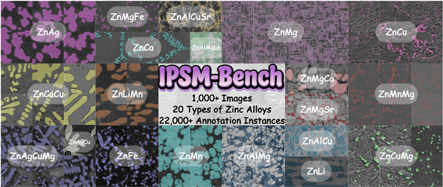
Therefore, we first construct a large-scale, high-quality annotated dataset of microstructural images of zinc alloys, named IPSM-Bench 1 , as shown in Figure 1. IPSM-Bench contains 1,054 microstructural images (512×512), comprising SEM and OM images, covering 20 types of zinc-based alloys and 22,179 manually annotated intermediate phase instances. All annotations are performed by experts in metallic materials science under quality control, ensuring precise delineation of intermediate phase boundaries. This dataset addresses a critical gap in the field by providing scarce, highquality annotated data that serves as a reliable foundation for training and evaluating intermediate phase segmentation models. Furthermore, we propose a new Spatial Context Prior-guided SAM method, named SCoP-SAM, which extracts intermediate phase gradient structure and grayscale property priors as spatial context and integrates them into both the encoder and decoder of the SAM (Segment Anything Model) to provide prior-aware prompts for intermediate phase regions. Our SCoP-SAM precisely anchors the target intermediate phase regions during segmentation and fully delineates their complex boundary contours, thereby significantly improving segmentation accuracy.
| Dataset | Material | Available | Anno.Type | Resolution | Alloy Types | Number of Images | Data Modality | |
|---|---|---|---|---|---|---|---|---|
| SEM | OM | |||||||
| NBS [Stuckner et al., 2022] | Ni-based Superalloy | Public | Mask | 512×512 | Unknown | 23 | � | � |
| UHCS-Seg [DeCost et al., 2019] | Ultrahigh Carbon Steel | Public | Mask | 645×475 | 1 | 24 | � | � |
| DP590-1 [Li et al., 2025] | Dual-phase Steel | Private | Mask | 512×512 | 1 | 23 | � | � |
| DP590-2 [Li et al., 2025] | Dual-phase Steel | Private | Mask | 1024×768 | 1 | 21 | � | � |
| Carbide [Ma and Yu, 2023] | Fe-0.2C-1.35Mn-2.5Cr-1.5Si Alloy | Private | Mask | 2048×1536 | 1 | 64 | � | � |
| IPSM-Bench (Ours) | Zinc-based Alloys | This Paper | Mask | 512×512 | 20 | 1,054 | � | � |
The main contributions are summarized as follows: 1) To the best of our knowledge, we construct the first large-scale, diverse, and extensively annotated high-quality benchmark IPSM-Bench for intermediate phase segmentation in zinc alloy microstructures, providing a reliable foundation for training intermediate phase segmentation models and a fair comparison platform to drive methodological advances in analyzing zinc alloy microstructures. 2) We propose a new spatial context prior-guided SAM method (SCoP-SAM) that incorporates gradient-structure and grayscale-property priors into SAM to address key challenges in intermediate phase segmentation-low contrast, difficulty detecting small targets, and complex morphologies. 3) Extensive experiments are conducted on the proposed IPSM-Bench benchmark dataset and two other representative public alloy datasets to demonstrate the effectiveness of our SCoP-SAM method for intermediate phase segmentation in microstructure images.
1 https://github.com/AgileMotionTeam/IPSM-Bench
2 RelatedWork
2.1 Metallic Microstructure Image Datasets
In the field of metallic microstructure image analysis, openaccess, large, and finely annotated datasets for intermediate phases remain scarce. As shown in Table 1, NBS [Stuckner et al., 2022] comprised only 23 intermediate phase particle images of nickel-based superalloys, with three segmentation classes: matrix phase, secondary precipitates, and tertiary precipitates. UHCS [DeCost et al., 2017] focused on the pearlite-to-spheroidized cementite transformation in ultrahigh carbon steel but lacked pixel-level annotations; its segmented variant, UHCS-Seg [DeCost et al., 2019], provided 24 images of intermediate phase particles. MetalDAM [Luengo et al., 2022] contained 42 high-resolution SEM images of additively manufactured steel, with pixel-level annotations for five microstructural components; however, intermediate phases accounted for an extremely low proportion (about 0.24%), leading to incomplete annotations that hindered the training of intermediate phase segmentation models. Notably, recent work [Li et al., 2025] has leveraged SAM for trainingfree microstructure analysis, but no standardized intermediate phase dataset has been established to date. Thus, there is an urgent need to develop a larger, more diverse, and more thoroughly annotated high-quality dataset for training intermediate phase segmentation models and as a fair comparison platform to advance research in this field.
2.2 SAM-based Image SegmentationModels in Multidisciplinary Applications
SAM [Kirillov et al., 2023], a foundational segmentation model based on the prompt-learning paradigm, enables zeroshot segmentation and supports multiple prompt types, improving the accuracy of complex-structure analysis [Ma et al., 2025; Abebe et al., 2025; Guru et al., 2025; Ma et al., 2024; Marks et al., 2025; Chen et al., 2024; Wu et al., 2025; Ji et al., 2024; Wu and Liu, 2026]. Recently, SAM-Adapter [Chen et al., 2023] developed a lightweight adaptation framework that converts task-specific knowledge into visual prompts to address poor generalization in specialized segmentation tasks. CAT-SAM [Xiao et al., 2024] proposed a few-shot conditional tuning network via a prompt-bridging structure that enables decoder-guided collaborative tuning of the encoder to address SAM's adaptation challenges. SAMCT [Lin et al., 2025] was equipped with a U-shaped CNN image encoder based on SAM, which provides supplementary local features for segmentation. DenseSAM [Zhou et al., 2025] replaced SAM's reliance on positional prompts in dense scenarios with semantic guidance, introducing an efficient semantic injection module and a dual-head decoding structure to tackle the challenge of dense objects in pathology and remote sensing images. MatSAM [Li et al., 2025] developed a SAM-based automatic analysis model for material microstructures by integrating a prompt-generation strategy and multi-scale feature fusion, enabling efficient segmentation of structures such as grains and second-phase particles. µSAM [Archit et al., 2025] provided a microscopy image segmentation and tracking tool based on SAM, unifying interactive and automatic segmentation across 2D/3D/temporal sequence data. Despite SAM's efforts in microstructure analysis, existing methods still suffer from recognition accuracy for fine structures (e.g., grains, precipitates, microcracks) and a high susceptibility to misjudgment in complex phase-distribution scenarios.
3 Dataset: IPSM-Bench
3.1 Construction Necessity
Zinc-based absorbable biomaterials exhibit unique advantages and broad application prospects in medical fields, owing to a moderate degradation rate matching the repair cycle of human bone tissue, excellent biocompatibility, and favorable mechanical properties [Li et al., 2020]. Therefore, we construct a new microstructure image dataset, named IPSMBench, spanning diverse zinc-based alloys to precisely analyze intermediate phases, which are critical for optimizing zinc alloy performance.
3.2 Data Acquisition
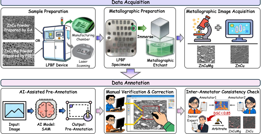
Our IPSM-Bench comprises 1,054 images, including 800 self-constructed and 254 sourced from the academic literature, covering two image types acquired via Optical Microscopy (OM) and Scanning Electron Microscopy (SEM). The data acquisition process is illustrated in Figure 2, which includes sample preparation, metallographic preparation, and metallographic image acquisition.
Sample Preparation. All samples were fabricated using the Laser Powder Bed Fusion (LPBF) technique. The raw materials were spherical zinc alloy powders produced by Gas Atomization (GA) and the Plasma Rotating Electrode Process (PREP). Fabrication was performed on a commercial LPBF system (SLM 125HL, SLM Solutions, Germany). Bulk solid samples were prepared by precisely adjusting laser processing parameters, with the laser energy density strictly maintained at 30-120 J/mm3 . Ultimately, bulk samples measuring 10×10×10mm were obtained. After removing the surface oxide layer, the samples' densities were measured using the Archimedes method. Only samples with densities above 99% were selected for subsequent metallographic preparation.
Metallographic Preparation. The surface parallel to the substrate was chosen for metallographic observation. Samples were subjected to a graded mechanical grinding process using 400#, 800#, 1500#, 3000#, and 5000# abrasive papers in sequence (#: grit of the sandpaper). After grinding, the surface was polished sequentially with W2.5µm, W1.5µm, and W0.5µm diamond metallographic polishing pastes until a bright, scratch-free surface suitable for microscopic observation was obtained. The polished observation surface was immersed in a dedicated enhanced metallographic etchant for 1-2 seconds, immediately removed, thoroughly rinsed with anhydrous ethanol, and dried prior to subsequent observation. Metallographic Image Acquisition. OM images of the microstructures are acquired using an optical microscope (BX53m, Olympus, Japan), while SEM images are obtained with a cold-field emission scanning electron microscope (SU8100, Hitachi, Japan). In addition to the experimentally acquired images, literature-derived data were collected by downloading microstructure images reported in relevant academic literature. These images were further processed by removing interfering regions and retaining valid areas to ensure consistency and usability within the dataset.
3.3 Data Annotation
To ensure annotation efficiency, we adopt an AI-assisted annotation strategy in this work. The entire annotation process comprises three key stages: AI model-based pre-annotation, manual verification and correction, and an inter-annotator consistency check.
AI-Assisted Pre-Annotation. A pre-trained segmentation model is used to generate initial segmentation masks for all images, thereby reducing the manual annotation workload. For zinc alloy intermediate phases with complex morphologies (e.g., lamellar, spherical, dendritic, and needle-like), SAM is selected as the AI auxiliary tool.
Manual Verification and Correction. We invite two professional annotators with more than 5 years of experience analyzing the microstructures of metallic materials to manually verify and correct the AI-generated pre-annotations. The correction criteria are based on the metallographic characteristics of zinc alloys: 1) Boundary Correction: For intermediate phases with blurred or inaccurate segmentation boundaries, the annotators adjusted the mask edges using professional annotation software (GIMP) to ensure precise delineation of intermediate phase contours. 2) Missing and False Annotation Correction: The annotators supplemented the missing smallscale intermediate phases that the AI model did not recognize and eliminated false annotations of matrix regions misidentified as intermediate phases.
Inter-Annotator Consistency Check. To ensure the reliability of the annotated data, an inter-annotator consistency check was conducted after manual correction. The Dice Similarity Coefficient (DSC) is used as an evaluation metric to quantify the consistency between the two annotators' results. For annotations with DSC<0.85 (i.e., low consistency), a third senior materials science expert was invited to arbitrate and determine the final annotation. The average DSC of the final annotated dataset exceeds 0.90, indicating high annotation consistency and reliability.
3.4 Statistics
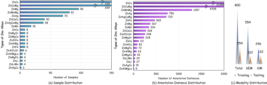
IPSM-Bench includes 20 Zinc-based alloy systems and comprises 1,054 images. Figure 3 (a) shows the distribution of Zinc alloy types, with ZnCu (370 samples, 35.10% of the total) and ZnCuMg (269 samples, 25.52% of the total) accounting for 60.63% of the total. IPSM-Bench has 22179 annotation instances. Figure 3 (b) illustrates the distribution of annotations across Zinc alloy types, with ZnCu (12355 annotation instances, 55.71% of the total) and ZnCuMg (4705 annotation instances, 21.21% of the total) accounting for 76.92% of the total. Figure 3 (c) presents the distribution of data modalities across training and testing. The training set includes 554 SEM images and 246 OM images, while the test set contains 122 SEM images and 132 OM images.
4 Methodology: SCoP-SAM
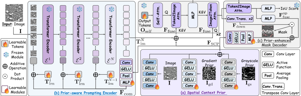
In this work, we propose a new Spatial Context Prior-guided SAM method (SCoP-SAM) for microstructure images. As shown in Figure 4, our SCoP-SAM comprises three core modules: Spatial Context Prior, Prior-aware Prompting Encoder, and Prior-enhanced Mask Decoder.
4.1 Spatial Context Prior (SCP)
To construct a spatial context prior for each input image, the SCP module integrates three types of information-RGB image, gradient prior, and grayscale prior-via three weightsharing-free learnable networks. Each network comprises three convolutional layers interleaved with two GELU nonlinearities. Specifically, given an input image I, we introduce three learnable networks fim, fgd, and fgs to compute the spatial context prior FSCP as follows:
where FSCP has a dimensionality of H1×W1×D1. Here, Igd (gradient prior) and Igs (grayscale prior) are derived via edge detection and grayscale thresholding algorithms, respectively. For the gradient prior, we employ an adaptive edge-detection algorithm to generate a gradient structure map, integrating the Canny operator and the Otsu thresholding method to extract edge features. Then, by comparing contour counts, the edge map with a greater number of contours is selected as Igd, aiming to maximize the preservation of topological integrity and structural continuity of the intermediate phase edges. For the grayscale prior, we model the grayscale distribution using dominant color analysis, yielding a grayscale map Igs. Specifically, we first analyze the distribution of black and white pixels in Iim and determine the target dominant color (black or white) based on the dominant pixel count. Subsequently, K-means clustering is applied to all image pixels, and the cluster center closest to the target dominant color is selected via Euclidean distance. Finally, a binary regional mask is generated around the cluster center, effectively capturing the aggregation properties of the intermediate phase region in the grayscale space.
4.2 Prior-aware Prompting Encoder (PPE)
Upon obtaining FSCP, we embed it into the subsequent Prioraware Prompting Encoder module to generate a robust prior prompt that guides downstream image encoding. Specifically, we introduce a learnable projection network fproj to project FSCP onto common prompting tokens Fcom. Subsequently, we add Fcom with Lp learnable tokens Tilrn to generate prioraware prompting tokens Tiprio for feeding into the i-th transformer encoder E i . This process can be formulated as:
where fproj is a learnable network consisting of a convolutional layer, GELU, average pooling, and an MLP. Notably, the PPE module incorporates N transformer encoders {E i }Ni=1 , where E i+1 encodes the output of E i , updating the prior-aware prompting token input fromTiprio to Ti+1 prio during encoding. For the final transformer encoder E N , we retain only TNim (i.e., the prior-enhanced image embedding tokens) and exclude TNprio from its output.
4.3 Prior-enhancedMask Decoder (PMD)
The PMD module integrates a spatial context prior and maps the prior-aware, token-guided image embedding to an output mask. Specifically, after obtaining the image embedding TNim , we add the spatial context prior FSCP to it and feed their integration Ffuse into a two-block decoder D1. Each block of D1 contains a self-attention layer, a cross-attention (token-toimage) layer, an MLP, and a cross-attention (image-to-token)
| Method | Venue | Backbone | IPSM-Bench (%) | ||
|---|---|---|---|---|---|
| mIoU | mF1 | mPre | |||
| SAM [Kirillov et al., 2023] | ICCV'23 | SAM ViT-H | 29.05 | 44.83 | 60.98 |
| SAM-Adapter [Chen et al., 2023] | ICCVW'23 | SAM ViT-H | 55.12 | 63.50 | 70.49 |
| SAMCT [Lin et al., 2025] | ICLR'24 | SAM ViT-H | 62.01 | 69.14 | 71.13 |
| CAT-SAM [Xiao et al., 2024] | ECCV'24 | SAM ViT-H | 43.05 | 55.60 | 34.87 |
| DenseSAM [Zhou et al., 2025] | IJCAI'25 | SAM ViT-H | 59.82 | 73.13 | 84.79 |
| MatSAM [Li et al., 2025] | Acta Materialia'25 | SAM ViT-H | 29.01 | 44.78 | 61.02 |
| µSAM [Archit et al., 2025] | Nature Methods'25 | SAM ViT-H | 39.19 | 44.08 | 42.48 |
| SCoP-SAM (Ours) | This Paper | SAM ViT-H | 71.52 | 82.82 | 87.55 |
| Method | Venue | NBS (%) | UHCS (%) | ||||
|---|---|---|---|---|---|---|---|
| mIoU | mF1 | mPre | mIoU | mF1 | mPre | ||
| SAM [Kirillov et al., 2023] | ICCV'23 | 62.54 | 76.86 | 79.11 | 32.00 | 45.66 | 52.60 |
| SAM-Adapter [Chen et al., 2023] | ICCVW'23 | 26.82 | 36.78 | 63.24 | 44.40 | 48.46 | 66.75 |
| SAMCT [Lin et al., 2025] | ICLR'24 | 34.50 | 43.80 | 49.19 | 43.55 | 46.62 | 90.50 |
| CAT-SAM [Xiao et al., 2024] | ECCV'24 | 24.73 | 33.50 | 55.12 | 43.48 | 46.51 | 46.01 |
| DenseSAM [Zhou et al., 2025] | IJCAI'25 | 29.18 | 40.78 | 74.18 | 37.59 | 48.36 | 49.94 |
| MatSAM [Li et al., 2025] | Acta Materialia'25 | 62.73 | 77.00 | 79.36 | 33.04 | 47.15 | 54.34 |
| µSAM [Archit et al., 2025] | Nature Methods'25 | 31.10 | 41.38 | 46.76 | 43.48 | 46.51 | 43.48 |
| SCoP-SAM (Ours) | This Paper | 67.26 | 80.42 | 80.43 | 57.25 | 67.48 | 80.93 |
Table 3: Comparison results against state-of-the-art methods on NBS and UHCS. The best and second-best results are in bold and underlined.
layer. A set of learnable output tokens Oself undergoes selfattention and serves as a query to attend to the prior-enhanced image embedding Ffuse, enabling it to capture global image context. Ffuse acts as the key and value for the token-to-image cross-attention, and the refined output token Ore self is updated via an MLP to integrate image contextual information. Ffuse serves as a query to attend to the updated output token via the image-to-token cross-attention, injecting global guidance into local spatial features and obtaining the prior-enhanced image embedding Fen fuse . After a two-block decoder D1, Fen fuse is upscaled via two transposed convolutions with GELU activations and layer normalization, yielding a downscaled feature map Fdown relative to the input image. The refined output token Ore self is fed into a token-to-image cross-attention layer, followed by an MLP that produces an upscaled image embedding Fup fuse . The point-wise sum of Fup fuse and Fcom (i.e., fcom(FSCP)) followed by the point-wise multiplication of Fdown and Fcom yields the foreground probability for each pixel, producing the final segmentation mask.
4.4 Training
The overall training objective of SCoP-SAM is formulated as a linearly weighted multi-task loss function that integrates three functionally complementary core loss components: a region-overlap optimization component (Dice loss), a pixelwise binary classification component (binary cross-entropy loss), and an auxiliary intersection-over-union (IoU) regression component for segmentation consistency.
5 Experiments
5.1 Experimental Setup
Datasets. To evaluate the proposed SCoP-SAM, we conduct experiments on various datasets, including IPSM-Bench, NBS [Stuckner et al., 2022], and UHCS [Na et al., 2023]. All methods are trained on the IPSM-Bench training set and evaluated on the test sets of IPSM-Bench, NBS, and UHCS. This design is due to the training sets for NBS and UHCS being too small to support model training. Specifically, for IPSMBench, the training set comprises 800 samples: 246 from the OM modality and 554 from the SEM modality, while the test set comprises 254 samples: 132 from the OM modality and 122 from the SEM modality. More details are provided in Figure 3 and subsection 3.4. UHCS has 5 labeled SEM images of cementite particles for testing. NBS has 9 labeled SEM images of nisuperalloy for testing.
Metrics. Following prior efforts [Zhou et al., 2025; Li et al., 2025; Chen et al., 2023], we adopt three pixel-level metrics: Intersection over Union (IoU), F1 score, and Precision. Specifically, we compute the performance for the foreground and background classes separately for each metric and report the mean across two classes, denoted mIoU, mF1, and mPre. Implementation Details. To ensure the fairness of comparative experiments, all experiments adhere to consistent configurations: 1) all methods use SAM ViT-H [Kirillov et al., 2023] as the backbone; 2) all methods use their official opensource implementations and settings, trained from scratch on IPSM-Bench's training set. We train SCoP-SAM on four NVIDIA A800 GPUs with a batch size of 4 for 50 epochs using the AdamW optimizer and a learning rate of 1e-4.
5.2 Results and Analysis
Performance Comparisons on IPSM-Bench. We compare our SCoP-SAM with SOTA methods [Zhou et al., 2025; Xiao et al., 2024; Chen et al., 2023; Lin et al., 2025; Archit et al., 2025; Kirillov et al., 2023; Li et al., 2025] on IPSM-Bench. In Table 2, SCoP-SAM consistently outperforms all methods. For instance, SCoP-SAM improves mIoU by 11.70% over DenseSAM, 9.51% over SAMCT, and 28.47% over CAT-SAM. We attribute these improvements primarily to incorporating a spatial context prior and prioraware prompts into the learnable modules of both the encoder and decoder, enabling robust performance under the challenges of intermediate phase segmentation, including low inter-phase contrast, blurred or incomplete boundaries, a high proportion of small-scale intermediate phases, heterogeneous morphologies, and interference from noise.
Performance Comparisons on NBS and UHCS. We further investigate the generalization of SCoP-SAM in few-shot settings with pronounced distribution shifts in materials data, such as large variations in precipitate size and more drastic morphological changes. To this end, we evaluate our SCoP-SAM on the more challenging NBS dataset and report the results in Table 3. Notably, the original SAM already achieves 62.54% on NBS, whereas several SAM-based variants (i.e., SAM-Adapter, SAMCT, CAT-SAM, DenseSAM, and µSAM) exhibit performance degradation, suggesting that their generalization under severe domain shifts remains limited. In contrast, SCoP-SAM outperforms MatSAM by 4.53%, further demonstrating the robustness of our method beyond the training data distribution. We attribute this advantage primarily to SCoP-SAM's use of multi-source spatial context priors to better exploit structural cues, enabling more stable object localization and boundary delineation under large-scale variations and morphological heterogeneity, thereby mitigating performance drops caused by few-shot learning and cross-domain distribution discrepancies. For the UHCS dataset, SCoP-SAM outperforms DenseSAM, SAMAdapter, and SAMCT by 19.66%, 12.85%, and 13.70% in mIoU, respectively, and demonstrates strong generalization, indicating that our IPSM-Bench also covers diverse intermediate phase morphologies and imaging conditions with high representativeness. Notably, SAMCT achieves a higher mPre (90.50%) than SCoP-SAM (80.93%) on UHCS; yet it fails to achieve a higher mIoU. This is because SAMCT has a higher miss rate (i.e., false negatives), predicting only a small set of high-confidence regions, which inflates precision, but insufficient mask coverage substantially degrades IoU.
| Module | mIoU (%) | mF1 (%) | mPre (%) |
|---|---|---|---|
| Baseline (ViT-H) | 29.05 | 44.83 | 60.98 |
| Baseline+SCP | 48.18 | 61.72 | 69.98 |
| Baseline+SCP+PPE | 59.49 | 72.98 | 81.38 |
| Baseline+SCP+PPE+PMD | 71.52 | 82.82 | 87.55 |
5.3 Ablation Study
We evaluate the effects of three core modules (i.e., SCP, PPE, and PMD). In Table 4, SCP alone achieves 48.18% mIoU; adding PPE improves performance to 59.49%; integrating PMD into the complete model yields optimal performance of 71.52%. These results demonstrate that each module positively contributes to segmentation performance and that the modules work synergistically to enhance overall performance, supporting SCoP-SAM's performance advantage.
5.4 Visualization Analysis
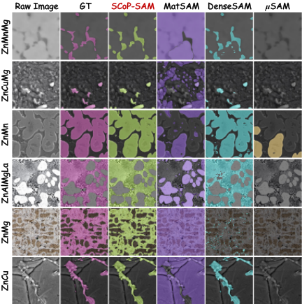
We provide a visual analysis of SEM and OM images of six typical zinc alloys, as shown in Figure 5. The SEM and OM images present distinct challenges: SEM images suffer from low contrast and background noise, while OM images exhibit complex topological structures of microstructural phases. Compared with the problems of over-segmentation, undersegmentation, or even complete failure in MatSAM, DenseSAM, and µSAM, our SCoP-SAM can stably and accurately restore irregular morphology and detailed boundaries, achieving high consistency with ground-truth (GT) annotations across all samples and demonstrating excellent crossmaterial and cross-modality generalization and robustness.
6 Conclusion
In this paper, we present IPSM-Bench, a new, large-scale, diverse, and high-quality benchmark for intermediate phase segmentation in zinc alloy microstructures, providing a reliable foundation for training and testing segmentation models. We further propose SCoP-SAM, which incorporates spatial context priors into image encoding and mask decoding generation, enabling more reliable use of the gradient structure and grayscale properties while improving both segmentation performance and boundary quality. Extensive experiments demonstrate that SCoP-SAM achieves state-of-the-art results on IPSM-Bench and delivers clear gains on two other public alloy benchmarks, indicating strong segmentation ability across various alloy scenarios.
Acknowledgments
This work was supported by grants from the National Natural Science Foundation of China (52541014, 52541013, 62522102, 52522110, 62432001), Beijing Natural Science Foundation (4252020, L247006), Fundamental Research Funds for the Central Universities (FRF-EF-25-05A), and Young Elite Scientists Sponsorship Program of the Beijing High Innovation Plan (20250747).
@inproceedings{ijcai2026p215,
  title     = {IPSM-Bench: A New Intermediate Phase Segmentation Benchmark in Microstructure Images of Zinc-Based Absorbable Biomaterials},
  author    = {Xu, Jinglin and Zhao, Shangyan and Wang, Jiabo and Mu, Xinghong and Lei, Yulong and Zhang, Jiacheng and Sun, Hongbo and Li, Yageng},
  booktitle = {Proceedings of the Thirty-Fifth International Joint Conference on
               Artificial Intelligence, {IJCAI-26}},
  publisher = {International Joint Conferences on Artificial Intelligence Organization},
  editor    = {Diego Calvanese},
  pages     = {1929--1937},
  year      = {2026},
  month     = {8},
  note      = {Main Track},
  doi       = {10.24963/ijcai.2026/215},
  url       = {https://doi.org/10.24963/ijcai.2026/215},
}
TY - CPAPER TI - IPSM-Bench: A New Intermediate Phase Segmentation Benchmark in Microstructure Images of Zinc-Based Absorbable Biomaterials AU - Xu, Jinglin AU - Zhao, Shangyan AU - Wang, Jiabo AU - Mu, Xinghong AU - Lei, Yulong AU - Zhang, Jiacheng AU - Sun, Hongbo AU - Li, Yageng T2 - Proceedings of the Thirty-Fifth International Joint Conference on Artificial Intelligence, IJCAI-26 PB - International Joint Conferences on Artificial Intelligence Organization PY - 2026 SP - 1929 EP - 1937 DO - 10.24963/ijcai.2026/215 UR - https://doi.org/10.24963/ijcai.2026/215 ER -Copied
Machine-readable versions of this paper, for tools and AI agents:





