
On AI-Assisted Pneumoconiosis Detection from Chest X-rays
Proceedings of the Thirty-Second International Joint Conference on Artificial Intelligence. AI for Good - Projects. Pages 6353-6361.
https://doi.org/10.24963/ijcai.2023/705
Keywords
- AI for Good - Projects: Machine Learning
- AI for Good - Projects: Multidisciplinary Topics and Applications
- AI for Good - Projects: Computer Vision
- AI for Good - Projects: Humans and AI
- AI for Good - Projects: AI Ethics, Trust, Fairness
Abstract
According to theWorld Health Organization, Pneumoconiosis
affects millions of workers globally,
with an estimated 260,000 deaths annually. The
burden of Pneumoconiosis is particularly high in
low-income countries, where occupational safety
standards are often inadequate, and the prevalence
of the disease is increasing rapidly. The reduced
availability of expert medical care in rural areas,
where these diseases are more prevalent, further
adds to the delayed screening and unfavourable outcomes
of the disease. This paper aims to highlight
the urgent need for early screening and detection
of Pneumoconiosis, given its significant impact on
affected individuals, their families, and societies as
a whole. With the help of low-cost machine learning
models, early screening, detection, and prevention
of Pneumoconiosis can help reduce healthcare
costs, particularly in low-income countries. In this
direction, this research focuses on designing AI solutions
for detecting different kinds of Pneumoconiosis
from chest X-ray data. This will contribute
to the Sustainable Development Goal 3 of ensuring
healthy lives and promoting well-being for all at all
ages, and present the framework for data collection
and algorithm for detecting Pneumoconiosis
for early screening. The baseline results show that
the existing algorithms are unable to address this
challenge. Therefore, it is our assertion that this
research will improve state-of-the-art algorithms of
segmentation, semantic segmentation, and classification
not only for this disease but in general medical
image analysis literature.
- [Akhter et al., 2023] Yasmeena Akhter, Richa Singh, and Mayank Vatsa. AI-based radiodiagnosis using chest x- rays: A review. Frontiers in Big Data, 6, 2023.
- [Behera et al., 2020] D Behera, VP Pannu, and Ranjan Ku- mar Behera. National tb elimination programme-can it end tb in india by 2025: An appraisal. Indian J Chest Dis Allied Sci, 62:203-27, 2020.
- [Bell and Mazurek, 2020] Jessica L Bell and Jacek M Mazurek. Trends in pneumoconiosis deaths-united states, 1999-2018. Morbidity and Mortality Weekly Re- port, 69(23):693, 2020.
- [Calvert et al., 2003] GM Calvert, FL Rice, JM Boiano, JW Sheehy, and WT Sanderson. Occupational silica expo- sure and risk of various diseases: an analysis using death certificates from 27 states of the united states. Occupa- tional and environmental medicine, 60(2):122-129, 2003.
- [Devnath et al., 2021] Liton Devnath, Suhuai Luo, Peter Summons, and Dadong Wang. Automated detection of pneumoconiosis with multilevel deep features learned from chest x-ray radiographs. Computers in Biology and Medicine, 129:104125, 2021.
- [Dwork et al., 2012] Cynthia Dwork, Moritz Hardt, Toniann Pitassi, Omer Reingold, and Richard S. Zemel. Fairness through awareness. In Shafi Goldwasser, editor, Innova- tions in Theoretical Computer Science 2012, Cambridge, MA, USA, January 8-10, 2012, pages 214-226. ACM, 2012.
- [Ehrlich et al., 2021] Rodney Ehrlich, Paula Akugizibwe, Nandi Siegfried, and David Rees. The association between silica exposure, silicosis and tuberculosis: a systematic re- view and meta-analysis. BMC Public Health, 21(1):953, 2021.
- [Gong et al., 2020] Sixue Gong, Xiaoming Liu, and Anil K. Jain. Jointly de-biasing face recognition and demographic attribute estimation. In ECCV (29), volume 12374 of Lec- ture Notes in Computer Science, pages 330-347. Springer, 2020.
- [He et al., 2016] Kaiming He, Xiangyu Zhang, Shaoqing Ren, and Jian Sun. Deep residual learning for image recog- nition. In 2016 IEEE Conference on Computer Vision and Pattern Recognition (CVPR), pages 770-778, 2016.
- [He et al., 2017] Kaiming He, Georgia Gkioxari, Piotr Dollár, and Ross Girshick. Mask R-CNN. In 2017 IEEE International Conference on Computer Vision (ICCV), pages 2980-2988, 2017.
- [Hosoda, 2005] Yutaka Hosoda. ILO international classifi- cations of radiographs of pneumoconioses-past, present and future-. In International Classification of HRCT for Occupational and Environmental Respiratory Diseases, pages 57-70. Springer, 2005.
- [Huang et al., 2017] Gao Huang, Zhuang Liu, Laurens Van Der Maaten, and Kilian Q. Weinberger. Densely con- nected convolutional networks. In IEEE Conference on Computer Vision and Pattern Recognition (CVPR), pages 2261-2269, 2017.
- [Maboso et al., 2023] Botembetume Maboso, Jim te Wa- ter Naude, David Rees, Hillel Goodman, and Rodney Ehrlich. Difficulties in distinguishing silicosis and pul- monary tuberculosis in silica-exposed gold miners: A report of four cases. American Journal of Industrial Medicine, 2023.
- [Mandal, 1988] AS Mandal. Pneumoconiosis in indian mines. East End Medical Publishers, Vol.c, 1988.
- [Poinen-Rughooputh et al., 2016] Satiavani Poinen- Rughooputh, Mahesh Shumsher Rughooputh, Yanjun Guo, Yi Rong, and Weihong Chen. Occupational expo- sure to silica dust and risk of lung cancer: an updated meta-analysis of epidemiological studies. BMC public health, 16(1):1-17, 2016.
- [Qi et al., 2021] Xian-Mei Qi, Ya Luo, Mei-Yue Song, Ying Liu, Ting Shu, Ying Liu, Jun-Ling Pang, Jing Wang, and Chen Wang. Pneumoconiosis: current status and future prospects. Chinese Medical Journal, 134(08):898-907, 2021.
- [Radford et al., 2018] Alec Radford, Karthik Narasimhan, Tim Salimans, and Ilya Sutskever. Improving language understanding by generative pre-training. OpenAI https:// cdn.openai.com/research-covers/language-unsupervised/ language understanding paper.pdf, 2018. Accessed: 2023-05-24. Link
- [Rajpurkar et al., 2017] Pranav Rajpurkar, Jeremy Irvin, Kaylie Zhu, Brandon Yang, Hershel Mehta, Tony Duan, Daisy Ding, Aarti Bagul, Curtis Langlotz, Katie Shpan- skaya, et al. Chexnet: Radiologist-level pneumonia de- tection on chest x-rays with deep learning. arXiv preprint arXiv:1711.05225, 2017.
- [Rao, 1934] S Subba Rao. Silicosis in india. British Medical Journal, 1(3832):1094, 1934.
- [Selvaraju et al., 2017] Ramprasaath R. Selvaraju, Michael Cogswell, Abhishek Das, Ramakrishna Vedantam, Devi Parikh, and Dhruv Batra. Grad-cam: Visual explanations from deep networks via gradient-based localization. In 2017 IEEE International Conference on Computer Vision (ICCV), pages 618-626, 2017.
- [Szegedy et al., 2015] Christian Szegedy, Wei Liu, Yangqing Jia, Pierre Sermanet, Scott Reed, Dragomir Anguelov, Dumitru Erhan, Vincent Vanhoucke, and Andrew Rabi- novich. Going deeper with convolutions. In 2015 IEEE Conference on Computer Vision and Pattern Recognition (CVPR), pages 1-9, 2015.
- [Wang et al., 2020] Xiaohua Wang, Juezhao Yu, Qiao Zhu, Shuqiang Li, Zanmei Zhao, Bohan Yang, and Jiantao Pu. Potential of deep learning in assessing pneumoconiosis de- picted on digital chest radiography. Occupational and En- vironmental Medicine, 77(9):597-602, 2020.
- [Wang et al., 2021] Zheng Wang, Qingjun Qian, Jianfang Zhang, et al. Deep learning for computer-aided diagno- sis of pneumoconiosis. Research Square, May 2021.
- [Xu et al., 2010] Horace Xu, Xiaodong Tao, Ramasubrama- nian Sundararajan, Weizhong Yan, Pavan Annangi, Xi- wen Sun, and Ling Mao. Computer aided detection for pneumoconiosis screening on digital chest radiographs. In Proceedings of the Third International Workshop on Pul- monary Image Analysis, pages 129-138, 2010.
- [Yang et al., 2021] Fan Yang, Zhi-Ri Tang, Jing Chen, Min Tang, Shengchun Wang, Wanyin Qi, Chong Yao, Yuanyuan Yu, Yinan Guo, and Zekuan Yu. Pneumoconio- sis computer aided diagnosis system based on x-rays and deep learning. BMC Medical Imaging, 21(1):1-7, 2021.
- [Zhang et al., 2021] Liuzhuo Zhang, Ruichen Rong, Qiwei Li, Donghan M Yang, Bo Yao, Danni Luo, Xiong Zhang, Xianfeng Zhu, Jun Luo, Yongquan Liu, et al. A deep learning-based model for screening and staging pneumo- coniosis. Scientific reports, 11(1):1-7, 2021.
References were extracted automatically from the PDF and may contain errors.
Full text extracted automatically from the PDF; figures, tables and equations may differ. The PDF is the version of record.
1 Introduction
Industries play a vital role in the economic growth and development of the country. They provide employment opportunities, stimulate growth in related sectors, and contribute significantly to the country's Gross Domestic Product (GDP). It is predicted that the Indian economy will grow by around 7% in the upcoming years. This growth is expected to be fuelled by sectors such as infrastructure. The mining sector plays a critical role in providing the raw materials required to sustain the growth of these rapidly expanding industries. Rajasthan has the largest number of mining leases in India, with a total of 33,122 licenses 1 , comprising 189 leases for major minerals, 15,245 leases for minor minerals, and 17,688 quarry licenses. The majority of these leases are for sandstone mines and quarries that are part of the unorganized and small-scale sector. However, with the rise of mining industries, there has also been an increase in occupational hazards such as Pneumoconiosis.
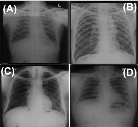
Pneumoconiosis is a group of interstitial lung disorders often found in people working in industries such as mining and construction. It is caused by prolonged exposure to substances such as silica, coal, asbestos, and mixed dust. This occupational disorder is irreversible and tends to get worse over time. It is a global problem becoming a leading cause of death among workers in certain industries. Pneumoconiosis can also be classified based on substances that are inhaled by workers, such as silica (Silicosis), cotton and other fibers
1 https://tinyurl.com/57b22pay
(brown lung), ash and dust (pneumonoultramicroscopicsilicovolcanoconiosis), asbestos (coal worker Pneumoconiosis or black lung), mixed-dust Pneumoconiosis (MDP), and Diacetyl (popcorn lung). People exposed to these substances are at a high risk of developing other lung problems, such as lung cancer, lung collapse, and TB [Ehrlich et al., 2021; Calvert et al., 2003; Poinen-Rughooputh et al., 2016]. Pneumoconiosis, in general, is rising in cases and is prevalent worldwide among industry workers [Qi et al., 2021; Bell and Mazurek, 2020].
Countless workers engaged in mines, construction, and factories silently die from exposure to these substances. The exact number of Pneumoconiosis patients worldwide is unknown because of a lack of proper reporting and poor workplace respiratory surveillance initiatives. In India, it is prevalent in the majority of states such as Gujarat, Rajasthan, Pondicherry, Haryana, Uttar Pradesh, Bihar, Chhattisgarh, Jharkhand, Orissa and West Bengal, with more than 10 million workers (including manufacturing, Mining, and construction sectors) at risk of industry dust exposure 2 . The prevalence of Pneumoconiosis in India ranges widely from 3.5% in ordnance factories to 54.6% in the slate-pencil industry [Behera et al., 2020]. This variation in prevalence is due to the substance concentrations in the different work environments, duration of exposure and job demands. The Government of India has started different initiatives to improve the life of workers at its risk; the Factory Act of India (1948) ordered the owners to provide well-ventilated working conditions, reduce the crowd, and provide provisions for protection from dust along with occupational health care. The state governments have also started portals to register workers for compensation. Moreover, the Occupational Safety, Health, and Working Condition Code 2020 (OSHWC) makes it mandatory for employers to provide annual health checks free of cost 3 . Several preventive measures have been initiated across places such as OSHA 4 , MSHA 5 and NIHOSH 6 by the Center for disease control and prevention. However, cases are still increasing and becoming the leading cause of death among occupational workers worldwide ([Qi et al., 2021; Bell and Mazurek, 2020]).
Pneumoconiosis confirmation diagnosis is generally performed using chest X-rays. While the findings of different Pneumoconiosis disorders become clearly visible with the progression of the disease; thus early screening of the diseases is a major challenge. The state-of-the-art algorithms are also primarily trained for tuberculosis prediction, and the inter-class similarities between the early stages of Pneumoconiosis and tuberculosis make it difficult to differentiate between them. In collaboration with local administration and radiologists, we have started working towards designing AIenabled solutions for detecting and early screening of Pneumoconiosis among Industry workers in India.
2 https://tinyurl.com/5y7fzkkx
3 https://tinyurl.com/4zwcy8uu
4 https://www.osha.gov/
5 https://www.msha.gov/
6 https://www.cdc.gov/niosh/index.htm
2 Description of the Task and Problem Statement
Pneumoconiosis is characterized by the symptoms such as long-term cough, shortness of breath, and phlegm. Medical examinations for diagnosing Pneumoconiosis include the collection of sputum samples or radiology-based methods such as chest X-rays and CT scans. The former takes more than 24 hours to find the results. On the other hand, radiologybased approaches are non-invasive and less time-consuming to diagnose the presence or absence of Pneumoconiosis. In some cases, a biopsy is also advised. Chest X-ray (CXR) is a practical tool recommended by the International Labour Organisation, ILO in 1958 [Hosoda, 2005] and is commonly available in different public health centres (PHCs). CXRs are an easy, non-invasive method to help detect pulmonary and interstitial disorders, including Pneumoconiosis. However, due to the spread of disease in specific regions where these industries are located, there is limited knowledge and expertise in Pneumoconiosis diagnosis among medical professionals, and there are chances of misdiagnosis of TB, which is also prevalent among workers [Ehrlich et al., 2021; Maboso et al., 2023]. This is due to the non-universal translation of Pneumoconiosis as a public health challenge. In addition, these industries are often located in remote regions, particularly in rural areas where healthcare is restricted to PHCs with limited availability of expert chest radiologists and pulmonologists. This further affects regular screening due to the low doctor-to-patient ratio.
Existing AI-based approaches for CXR-based image analysis have focused on the common pathologies present in the lungs. The existing work on pulmonary disorders focused on available datasets for TB, Pneumonia, cardiomegaly, lung cancer and many others. However, occupational diseases such as Pneumoconiosis are under-represented in the AI research community. Limited research in diagnosing Pneumoconiosis using CXRs is based on a small sample size and in-house datasets. [Akhter et al., 2023] reviewd the existing work done to detect Pneumoconiosis from CXRs highlighting the dataset as one of the main challenges. [Yang et al., 2021] collected CXRs from 2017 to 2020 and ended their study with 1760 samples. [Zhang et al., 2021] provided a deep learningbased algorithm for diagnosing and staging Pneumoconiosis for their in-house collected 1216 samples, including normal cases. Similarly, [Xu et al., 2010; Wang et al., 2020; Devnath et al., 2021; Wang et al., 2021] studied AI-based Pneumoconiosis detection on small sample inhouse datasets. None of these datasets is publicly available for designing interpretable deep learning-based algorithms for fast and accurate diagnosis of Pneumoconiosis.
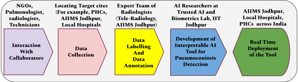
In this research, we are working in collaboration with the All India Institute of Medical Sciences in Jodhpur, India, Tele-radiology Bangalore, PHCs in Rajasthan and NGOs working in the direction of the upliftment of the industry worker population. The outcome of the work will be an AIbased detection tool, which can be used as an digital assistant, especially at the PHCs in rural areas of India. An automatic detection system available to hospitals can ease the diagnosis process and encourage early detection. Early detection of Pneumoconiosis, an irreversible and progressive occupational disorder, can greatly aid in improving the life expectancy of workers. The complete pipeline of the proposed work is showcased in Fig. 2.
3 Target SDG(s) and Societal Benefits
Pneumoconiosis disproportionately affects marginalized and vulnerable populations, such as workers in low-income countries or those who work in hazardous occupations without proper safety measures. Its detection and prevention are critical to ensure these workers are not left behind in pursuing better health outcomes. This aligns with the United Nations "Leave No One Behind" policy, a fundamental principle of the 2030 Agenda for Sustainable Development. In particular, SDG target 3.9 aims to reduce the number of deaths and illnesses caused by hazardous chemicals and air, water, and soil pollution and contamination. By improving the early detection of Pneumoconiosis, we can prevent further damage to workers' health and well-being, thus contributing to the achievement of this target. The UN policy recognizes that the SDGs can only be achieved if they focus on the equality and inclusion of low-income and middle-income countries. By prioritizing the detection of Pneumoconiosis in marginalized and vulnerable populations, we can help to ensure that these individuals receive the care and support they need to improve their health and well-being. This aligns with SDG target 10.2, which aims to empower and promote the social, economic, and political inclusion of all, irrespective of age, sex, disability, race, ethnicity, origin, religion, or economic status. The SDG target 3.3 aims to end the epidemics of AIDS, TB, malaria, and neglected tropical diseases and combat hepatitis, water-borne diseases, and other communicable diseases. With the early detection of Pneumoconiosis, we can help to prevent further TB transmission and promote public health. The early detection using the developed tool will help to achieve target 3.4, which aims to reduce mortality due to noncommunicable diseases. The impact of Pneumoconiosis is not limited to the individual worker. It can have far-reaching economic consequences for families and communities, especially when the worker is the primary breadwinner. By detecting Pneumoconiosis early, we can help to prevent the loss of income and the resulting poverty that can follow. This aligns with target 1.1, 1.4,1.5, which aims to eradicate extreme poverty and target 8.5, which seeks to achieve full and productive employment and decent work for all. The early detection of Pneumoconiosis is critical for achieving several of the UN-SDG, including health, poverty reduction, and decent work targets. By investing in technology and strategies to improve Pneumoconiosis detection, we can progress towards a more just, equitable, and sustainable world.
4 Challenges and Risks
Early diagnosis of Pneumoconiosis faces different challenges from an AI and computer vision perspective. The development of an AI-based tool for Pneumoconiosis detection involves addressing the following core research questions:
- Large Data Collection and Sharing: The problem of Pneumoconiosis detection is under-represented in the AI
and computer vision domain due to unavailable datasets. Accessing data for other lung disorders can be collected from any general hospital. However, data collection for automating Pneumoconiosis requires visiting the PHCs, and local hospitals and collaborating with NGOs to motivate the worker population to get tested and ready to share their working history. Covering the different industries such as stone crushing, glass production, coal processing, and many others is a challenging but an important process to enable the development of the AIbased Tool.
- Handling Interoperability: The proposed work aims to incorporate data from multiple industrial sites across India and work with the existing healthcare infrastructure. Therefore, it is important that the model works efficiently for the varied data acquisition setup that exists across different hospitals and primary healthcare centres. The variations are owed to the availability of multiple sensors, including X-ray Machines and digital scanners. Moreover, due to the lack of sophisticated infrastructure in rural settings, conventional screen film radiography is still persistent and commonly used compared to digital X-ray machines.
- Dataset Labelling and Annotation: Building an explainable AI algorithm for prediction requires detailed data labelling and annotation in terms of segmentation, findings, and disease from well-trained domain experts. Therefore, partnership domain experts, including radiologists and pulmonologists, is essential.
- Poor Quality and Noisy Samples: Given the largescale infrastructure and the large variations across operators and acquisition devices, there is a possibility of poor quality and noisy samples. Further, there are possibilities of data loss during communication as well. Therefore, the presence of noisy and poor-quality samples is another challenge that needs to be incorporated while designing the model.
- Lung disease correlation and co-occurrence: Pneumoconiosis patients also suffer from other lung disorders, such as TB and COPD, and show similar symptoms. Similarly, different correlated manifestations in the lungs can co-exist in the CXR sample and lead to misdiagnosis. Therefore, a cohort of experienced radiologists and pulmonologists is required to consider these factors for manual labelling to develop an efficient and accurate AI algorithm.
- Model Explainability: AI-based solutions for healthcare applications require developing interpretable and explainable solutions. This will ensure that the domain experts utilizing these solutions can verify and confirm the findings and the diagnosis.
- Model Deployment: Deploying detection tools has to go through compliance testing and approval from regulatory agencies. Moreover, since the application of this technology is primarily meant for low-income, low-infrastructure sites, a thorough consideration has to be given to the infrastructure requirements, including
power and network. Further, the latency should be quite low so to speed up the screening process.
5 Goals
The primary aim of this research is to develop an explainable and accurate AI-enabled solution for automatically predicting the presence of different types of Pneumoconiosis from the given input CXR images. The availability of such a tool can assist health practitioners in detecting the presence of abnormalities and predicting the occurrence of Pneumoconiosis in the lungs. As shown in Fig. 3, developing such a tool requires learning AI models from large and annotated datasets. Therefore, some of the intermediate objectives of the project are:
- Creating a large CXR-based dataset from the Indian hospitals and PHCs for different types of Pneumoconiosis,
- Annotating the dataset with complete global and local labelling by domain experts,
- Designing a unified model enabling the use of local labelling (findings) to achieve explainability and image data for disease detection and report generation, and
- Testing and verification of the model in real-world conditions.
We further aim to integrate this approach into a telemedicine service to enable usage in remote locations. While this work aims to detect and distinguish different variants of Pneumoconiosis in the Indian population, it can be further extended for other CXR-based medical investigations. We believe our work will encourage more collaboration of different stakeholders (NGOs, doctors, AI researchers, and Government officials) in achieving UN SDG goals 3 (Ensure healthy lives and promote well-being for all at all ages), 9 (Build resilient infrastructure, promote sustainable industrialization and foster innovation), and 10 (Reduce inequality within and among countries). We hope our work encourages more AI researchers across the globe to develop novel solutions for such socially relevant problems. The overall collaborative effort will provide a comprehensive approach by identifying the potential cases and providing a well-evaluated program for preventing, controlling, detecting, and eliminating this irreversible interstitial lung disorder.
6 Method
The proposed methodology for detecting and classifying different pathologies in CXR follows a sequential process comprising three primary steps: (1) preprocessing and segmentation, (2) finding the abnormalities, and (3) disease prediction. Figure 3 illustrates the flow diagram of the methodology.
Firstly, the preprocessing and segmentation module enhances the image quality and identifies the regions of interest. This step aims to improve the input image and accurately locate the regions of interest. Subsequently, the bounding box generation module utilizes deep learning to segment each pathology in the image, create a bounding box around it, and generate a local label. This module is trained on a dataset of CXR images with pathology labels and associated bounding boxes. Mask R-CNN [He et al., 2017], a well-known architecture, can be employed to achieve this. By refining the initial proposals generated by the Region Proposal Network (RPN), Mask R-CNN can learn to generate bounding boxes for each pathology. These bounding boxes enable healthcare professionals to visualize the pathologies as local labels in the image, aiding in the diagnosis of Pneumoconiosis.
After generating the bounding boxes, the classification module comes into play. This module categorizes the diseases associated with the image on the global label. To accomplish this task, a deep neural network model is developed, leveraging the CheXNet architecture [Rajpurkar et al., 2017]. CheXNet is a convolutional neural network trained on a large dataset of CXR images with disease labels. By utilizing a pre-trained network and fine-tuning, the CheXNet model is adapted to the specific task of disease classification, alleviating the domain shift when training the classification network for Pneumoconiosis. The trained classification module extracts relevant features from the preprocessed image, which are then used to predict the presence or absence of each disease in the image as global labels.
Furthermore, to provide comprehensive medical reports, a Transformer architecture called GPT (Generative Pre-trained Transformer) [Radford et al., 2018] can be employed. GPT, which has been used for generating natural language text, which aids in generating medical reports based on the bounding boxes generated by Mask R-CNN. The GPT model is pretrained on extensive text data and fine-tuned for specific Natural Language Processing (NLP) tasks, such as generating reports from medical images. By incorporating local information from the findings, the generated reports ensure pre-hoc model explainability and deliver reliable results based on the diseased (local) labels. These reports can assist doctors in diagnosing pneumoconiosis effectively. Additionally, during the algorithm development, measures are taken to incorporate debiasing techniques, such as fair data acquisition and regularizing loss for subgroups (gender, age), to ensure fairness in the tool's output.
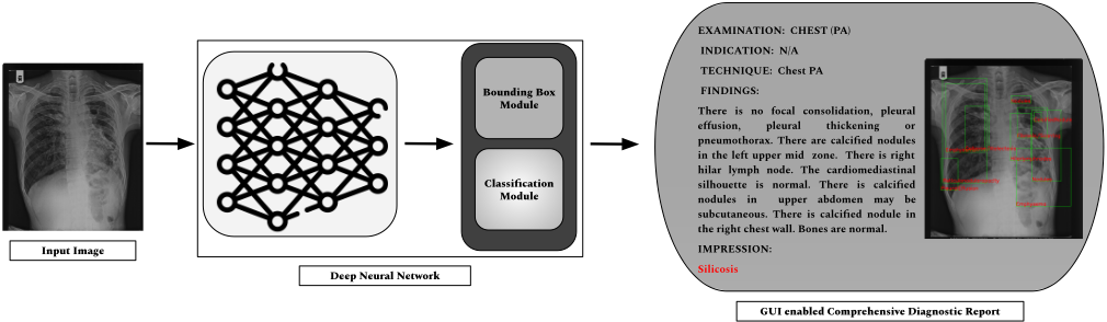
Throughout the deep learning architecture, various stages incorporate explainability to ensure transparent decisionmaking understandable by healthcare professionals. For instance, post-hoc visualizations and model-agnostic techniques, such as Grad-CAM [Selvaraju et al., 2017], can be utilized to generate explanations for the model's decision. Grad-CAM enables visualization of the regions in the input image that influenced the disease detection model's decision in generating the corresponding bounding box. By employing such holistic detection approaches encompassing model explainability and fairness, the proposed tool aims to provide comprehensive support in diagnosing pneumoconiosis.
7 Case Study
In this research, we undertake Silicosis, which is one of the major types of Pneumoconiosis, and it occurs due to the presence of Silica, SiO2. India reported its first Silicosis patient in 1934 [Rao, 1934], and its widespread effect is reported in India's long history from Kolar Gold Fields (KGF) where about 43.7% workers were found to be Silicosis affected [Mandal, 1988]. Silicosis is a serious and widespread occupational disease affecting workers in various industries, including stone carving, ceramics, mining, construction, mineral processing, tunneling, glass-making, foundries, and sandblasting. Given the significant impact of Silicosis on workers, their families, and the economy, there is a need for increased awareness of the disease and improved occupational health and safety standards in high-risk industries. In most developing and many developed nations, Silicosis is responsible for high worker morbidity and mortality. It results in progressive massive fibrosis and Pneumothorax, and long over-exposure results in pulmonary TB. Silicosis and TB develop an association, resulting in Silicotuberculosis and leading to 2.5 times failure in treating Pulmonary TB.
In this project, we plan to work with the Sirohi district in the state of Rajasthan. Sirohi is an aspirational district under the government's Aspirational Districts Programme (ADP), which aims to develop the most underdeveloped districts in the country. Different developmental programs under ADP focus on five broad socio-economic themes, including Health & Nutrition, Education, Agriculture & Water Resources, Financial Inclusion & Skill Development, and Infrastructure. This specific case study focuses on Health and Nutrition.
According to Rajasthan health records 7 , out of 4931 detected silicosis cases between 2013-2017, 449 people have died. However, the lack of infrastructure for diagnosis and certification has made it difficult to determine the exact number of silicosis patients. Recently, the Rajasthan government has started a focused program with an online portal to register persons for certification and disbursement of relief. Under this, the initial screening of the individuals is conducted at the Community Health Centre; if suspected, the case is referred to the district Pneumoconiosis board for certification. The district Pneumoconiosis board consisting of one radiologist, one chest physician, and one general physician examines the person in detail and, based on chest radiographs and other investigations, certifies Silicosis. The details are sent to district authorities for validation, followed by payment of relief to the victim from the centralized "Pneumoconiosis Fund" set up by contributions from the BOCWWelfare Board, District Mining Foundation Trusts (DMFT), and the state government. As of May 31, 2022, a total of 181,687 individuals have enrolled for the silicosis screening examination. Since the system became available online, the district Pneumoconiosis boards have certified 23,436 cases of Silicosis, including 6,876 cases of death due to Silicosis. Prior to the launch of the online system, 17,687 cases, including 1,857 deaths due to Silicosis, were certified until May 2019. Relief has been distributed to a total of 30,293 individuals affected by Silicosis, and 31,121 silicosis victims have registered for disability pensions. Although the exact amount is not available, it is estimated that more than 5.5 billion INR (US $ 73.3 million) have been paid to silicosis victims as a relief. The state government is also in the process of establishing a Project Monitoring Unit for policy implementation. It has allocated 0.05 billion INR (US $ 670,000) for research and prevention of Silicosis.
7 https://tinyurl.com/silicosiscag
| Model | Classes | F1-Score | Class-wise Accuracy | Class-wise AUC |
|---|---|---|---|---|
| ResNet18 [He et al., 2016] | Normal Tuberculosis Silicosis SilicoTuberculosis | 0.58 | 0.75 ± 0.010 0.46 ± 0.090 0.54 ± 0.010 0.64 ± 0.007 | 0.91 ± 0.001 0.75 ± 0.008 0.85 ± 0.005 0.87 ± 0.005 |
| DenseNet121 [Huang et al., 2017] | Normal Tuberculosis Silicosis SilicoTuberculosis | 0.54 | 0.76± 0.003 0.47 ± 0.160 0.52 ± 0.002 0.43 ± 0.020 | 0.90 ± 0.003 0.76 ±0.018 0.83 ± 0.005 0.84 ± 0.013 |
| InceptionV3 [Szegedy et al., 2015] | Normal Tuberculosis Silicosis SilicoTuberculosis | 0.60 | 0.74 ± 0.009 0.45 ± 0.010 0.53 ± 0.007 0.59± 0.017 | 0.87± 0.007 0.71 ± 0.004 0.81 ± 0.007 0.88 ± 0.006 |
As a part of our case study, we gathered a total of 4,077 CXR samples. These samples were obtained from the primary healthcare centers in Sirohi. The study included 2,334 male subjects aged between 16 and 60 years and 435 female subjects aged between 20 and 50 years of age. Radiologists from Tele-Radiology Solutions Bangalore annotated these samples with various disease labels such as Silicosis, Silicotuberculosis, tuberculosis, and Normal, and findings such as Atelectasis, Cardiomegaly, Consolidation, Edema, Enlarged cardiomediastinum, Fracture, Lung lesion, and Lung opacity. The dataset is divided into two separate subsets: Set A contains images with only disease labels, while Set B contains images with lung segmentation maps, annotations, and disease labels.
We performed a baseline evaluation of existing deep learning architectures on this dataset, and the results are summarized in Table 1. We evaluated the baseline models using F1score, classwise accuracy, and classwise AUC. It can be seen from the results that normal and Silicotuberculosis classes are well differentiated due to high dissimilarities from the other two classes. However, models have shown lower prediction performance for Silicosis and TB. The results signify that the undetected Silicosis result in the worsening of the lung tissue, and diseased regions become more prominent in Silicotuberculosis. This emphasizes the need for early diagnosis of Silicosis. We have also developed a user interface for health professionals to test as a proposed solution. We have incorporated feedback from health professionals into the architecture to ensure thorough testing before final deployment. Our collaboration between the Indian Institute of Technology, IIT Jodhpur, and AIIMS Jodhpur can potentially enhance people's living standards in the Sirohi district. We aim to develop an interpretable AI engine that can differentiate between TB and Silicosis, helping doctors make accurate diagnoses and provide appropriate treatment, improving our preliminary results. This case study is a great example of how social responsibility and scientific research can work together to make a positive change.
8 Evaluation Criteria
The proposed automatic detection tool for early detection of Pneumoconiosis from CXRs will be evaluated in terms of the existing evaluation metrics for performance, fairness, and latency at the inference time. Existing works for evaluating an automatic detection tool involve different metrics across the tasks in medical image analysis. As shown in Figure 3, the proposed method comprises of multiple tasks, such as detecting and localizing the diseased areas (findings or local labels) comprising bounding boxes and predicting the global class label. The former will be evaluated using mean Average precision, mAP and Intersection over union, IOU metrics. While the later will be evaluated using overall accuracy, Area under the curve (AUC), classwise accuracy, sensitivity, specificity, and F1-score at a 95% Confidence Interval. To evaluate the proposed method for fairness, we will use the Degree of Bias, DoB [Gong et al., 2020] and Group fairness [Dwork et al., 2012] as the evaluation metrics.
9 Expected Results and Long-Term Impact
Working in small factories and industry is the norm for both males and females in rural areas. People willingly agree to work with poor ventilation and unhygienic conditions at workplaces to earn a livelihood. They even are unregistered, being paid on a daily basis to claim the issued relief from the government. Prolonged exposure to different types of dust at the workplace drastically increases the chances of Pneumoconiosis. Revolutionizing the healthcare sector is a need in terms of accuracy, speed, and reliability to achieve the goals of the right to good health. AI has the potential to achieve these features, and deploying AI-Based tools lowers the demand for manpower in rural areas. The proposed work aims to develop an AI-based tool for Pneumoconiosis detection from CXRs with improved performance in terms of sensitivity and AUC with equal treatment for subgroups in the population.
A "Digital Diagnostic Assistant" at PHCs will facilitate smooth diagnostics and provide efficient and reliable predictions by learning the disease patterns and providing potential diagnostic decisions. Pneumoconiosis detection using CXRs is a challenging task involving differentiating related lung disorders such as TB. AI-based tools for Pneumoconiosis detection could change the traditional method and result in a complete paradigm shift and pave the path for its early detection and achieve UN SDG goal 3 targets 3.3, 3.4, 3.8, 3.9, and 3.d. Earlier detection will aid in improving the health of the worker and manage the monetary expenses for the treatment of Pneumoconiosis, achieving UNSDG 1 target 1.1, 1.4, 1.5 and 1.a, 10.1 and 10.2.
We hope that our work will encourage more inclusive collaboration of stakeholders such as NGOs, Policy makers, medical domain experts, AI researchers, and funding agencies. We believe most of the workers would be included and made aware of this hazardous health challenge, and the establishment of well-ventilated industries and factories and regular monitoring of the worker's health would be initiated. A collaborative effort will prevent, control and implement programs to eradicate this challenging healthcare problem.
10 CollaborationMechanism, Timeline and Outputs
This project aims to develop and deploy an explainable AI algorithm-based tool for the early detection of Pneumoconiosis from CXRs. Achieving this objective requires collaborating and utilizing the expertise of a varied range of disciplines, such as researchers with AI and computer vision skills for developing AI-based models, pulmonologists and radiologists with domain expertise, public health professionals for deployment and testing, and NGOs for encouraging and counselling of the worker population to get checked on a regular time interval, prior to the appearance of any symptoms. We are currently working on this project with these partners. The key milestones of the project are as follows:
- After following the due process of the ethics board, the dataset is being collected by IIT Jodhpur researchers from the PHCs in the districts of Rajasthan.
- After cleaning and basic organization, the collected CXR data is annotated by radiologists, with annotations being performed by multiple groups of doctors to minimize any inconsistencies. PHC doctors will cross-check a subset of the samples, and a consensus will be reached in consultation with radiologists.
- The researchers at IIT Jodhpur will develop an AI-based model by incorporating local and global label information to train deep learning-based architecture, ensuring model explainability and fairness. The deployed model at PHCs of Rajasthan and other hospitals will be rigorously tested to check for real-time Pneumoconiosis detection.
- A user-friendly interface will be developed for the AI algorithm, enabling medical professionals to input CXRs and receive the algorithm's results.
The scientific outcomes of the proposed project include the following:
- A new scientific research thread will be established to propose a novel AI-based approach for Pneumoconiosis detection with the release of datasets and trained models publicly.
- Database comprising samples of healthy individuals and different variants of Pneumoconiosis, which will further promote research in this area.
- Paper publications on different aspects of the project:
- (i) Dataset of CXR images from patients diagnosed with Pneumoconiosis and healthy individuals. The paper will discuss not only the characteristics of the dataset but also the ethical and social implications of collecting such data.
- (ii) AI algorithm architecture developed for the early detection of Pneumoconiosis from CXRs. and
- (iii) A publication that explores the social implications of the AI algorithm for the early detection of Pneumoconiosis, which can contribute to improving public health and inform policies and practices around the use of AI in medical diagnosis.
11 Ethical Considerations
The proposed work aims to address the socially relevant occupational disorder, Pneumoconiosis, among industrial workers. Its first primary step comprises collecting human data in the form of CXRs and associated metadata (age, gender, name, and address of the data collection centers) from humans. Therefore, a strict data privacy policy needs to be followed. We have received approval from the Institute Review Board (IRB) at IIT Jodhpur, India, to initiate the proposed work. The subjects will be informed apriori about the reason for data collection, a consent form will be filled out, and proper permission will be obtained from the authorities involving medical officers in hospitals. The collected data will be stored and shared anonymously for data annotation and will be completely de-identified for future release. Hence, no personal information about the subject (worker) will be collected or released. Access to personal data such as age and gender will only be used for analysis during the study. We also strictly adhere to the ethical standards of the IJCAI conference.
For the algorithmic part, we involve a collaborative effort of all the stakeholders, ensuring smooth progress of the work and avoiding any unethical mistake from development to deployment of the tool. We ensure that the tool will be evaluated for model fairness, leaving no room for discrimination against any subgroup. The final tool will be released free of cost to the PHCs and local hospitals after testing and receiving usage approval.
Acknowledgments
This research is supported through a grant from MEITY. M. Vatsa is partially supported through SwarnaJayanti Fellowship by the Government of India. The authors also acknowledge the medical practitioners for their help in sample collection.
A About the Authors
Yasmeena Akhter is a Ph.D. student at the Indian Institute of Technology (IIT) Jodhpur, India. Her research focuses on interpretable deep learning for healthcare applications. She serves as the reviewer of conferences such as NeurIPS, CVPR, and MICCAI and journals, including Pattern Recognition Journal and IEEE T-BIOM.
Rishabh Ranjan is a Ph.D. student at IIT Jodhpur, India. His research interests include audio forensics, interpretable deep learning, and medical imaging. He serves as a reviewer at top venues such as NeurIPS.
Mayank Vatsa is a Professor at the Department of Computer Science and Engineering at the Indian Institute of Technology (IIT) Jodhpur, India. He serves as Dean of Research and Development and Project Director of the Technology and Innovation Hub (TIH) at IIT Jodhpur, India. He is a Fellow of IEEE and IAPR and the recipient of the prestigious SwarnaJayanti Fellowship. He was a member of the Indian Biometrics Standards Committee for e-gov applications. He also served as a member of the UIDAI's Biometrics Standard Subcommittee member (face, fingerprint, and iris standards). He also served as the Vice President (Publications) for IEEE Biometrics Council, where he led the efforts to start IEEE Transactions on Biometrics, Behavior, and Identity Science. His areas of interest are computer vision, machine learning, biometrics, and Trusted AI.
Richa Singh is a Professor at the Department of CSE, IIT Jodhpur. She received a Ph.D. degree in computer science from West Virginia University, USA. She is a Fellow of IEEE and IAPR. She has received several awards, including the Kusum and Mohandas Pai Faculty Research Fellowship at the IIIT-Delhi, the FAST Award by the Department of Science and Technology, India, and several best paper and best poster awards in international conferences. She has also held leadership positions in professional organizations, such as Program Co-Chair of CVPR2022, General Chair of FG2021, Vice President - Publications of the IEEE Biometrics Council, and Associate Editor-in-Chief of Pattern Recognition.
Santanu Chaudhary is a Professor with the Department of Electrical Engineering, IIT Delhi, India. He is currently serving as the Director of IIT Jodhpur, India. Before joining IIT Jodhpur, he completed his tenure as the Director of the Central Electronics Engineering Research Institute, Pilani, India. He has authored or co-authored more than 300 research publications in peer-reviewed journals and conference proceedings, 15 patents, and four authored/edited books to his credit. His research interests include image and video processing, computer vision, machine learning, and embedded systems. He is a Fellow of the Indian National Academy of Engineering, the National Academy of Sciences, and the International Association for Pattern Recognition. He was a recipient of the Distinguished Alumnus Award from IIT Kharagpur, the Indian National Science Academy Medal for Young Scientists in 1993, and the Advanced Computing and Communications Society-Centre for Development and Advanced Computing (ACCS-CDAC) Award for his research contributions in 2012.
@inproceedings{ijcai2023p705,
  title     = {On AI-Assisted Pneumoconiosis Detection from Chest X-rays},
  author    = {Akhter, Yasmeena and Ranjan, Rishabh and Singh, Richa and Vatsa, Mayank and Chaudhury, Santanu},
  booktitle = {Proceedings of the Thirty-Second International Joint Conference on
               Artificial Intelligence, {IJCAI-23}},
  publisher = {International Joint Conferences on Artificial Intelligence Organization},
  editor    = {Edith Elkind},
  pages     = {6353--6361},
  year      = {2023},
  month     = {8},
  note      = {AI for Good - Projects},
  doi       = {10.24963/ijcai.2023/705},
  url       = {https://doi.org/10.24963/ijcai.2023/705},
}
TY - CPAPER TI - On AI-Assisted Pneumoconiosis Detection from Chest X-rays AU - Akhter, Yasmeena AU - Ranjan, Rishabh AU - Singh, Richa AU - Vatsa, Mayank AU - Chaudhury, Santanu T2 - Proceedings of the Thirty-Second International Joint Conference on Artificial Intelligence, IJCAI-23 PB - International Joint Conferences on Artificial Intelligence Organization PY - 2023 SP - 6353 EP - 6361 DO - 10.24963/ijcai.2023/705 UR - https://doi.org/10.24963/ijcai.2023/705 ER -Copied
Machine-readable versions of this paper, for tools and AI agents:





